Categories
Diseases

Amyloidosis

Amyloidosis with pulmonary involvement is rare and occurs in primary or secondary forms (e.g., with connective tissue disease). HRCT can be completely normal, but occasionally nodules, reticular pattern and consolidation may occur. Calcification of the lung parenchyma is common.

Categories
Diseases

Alveolar Microlithiasis

Alveolar microlithiasis is a very rare disease. On CT (Figure 135) diffuse micronodular calcifications with a sand-like appearance can be seen in the basal-dorsal regions. Typically, the condition exhibits a poor correlation between radiographic findings and symptoms, often the patient appears worse on CT than clinically.

Figure 135. Man in his fifties with known alveolar microlithiasis. CT shows a “sandstorm”-like picture with extensive small calcifications.
Categories
Diseases

Lipoid Pneumonia

Exogenous lipoid pneumonia occurs following inhalation of mineral oils or aspiration of certain animal or vegetable oils. Fatty acids accumulate in the lung parenchyma resulting in inflammation, which can cause focal fibrosis. Endogenous (idiopathic) lipoid pneumonia also occurs with accumulation of lipids in the alveoli. HRCT in both forms show a mixture of ground-glass, crazy paving and consolidation. Distribution may be unilateral or bilateral, and sometimes the condition remains relatively unchanged. There is no cure. Lipoid pneumonia is a rare disease and diagnosis is confirmed by biopsy (Figures 33, 134).

Figure 134. A and B. Middle-aged woman who developed biopsy-verified lipoid pneumonia. The changes consist of ground-glass and crazy paving with elements of consolidation. The CT-findings remained relatively unchanged for several years. This was diagnosed as endogenous lipoid pneumonia.

A.
B.

Figure 134. A and B. Middle-aged woman who developed biopsy-verified lipoid pneumonia. The changes consist of ground-glass and crazy paving with elements of consolidation. The CT-findings remained relatively unchanged for several years. This was diagnosed as endogenous lipoid pneumonia.

Categories
Diseases

Alveolar Proteinosis

Alveolar proteinosis (Figures 27, 133) is a rare lung disorder, whereby the alveoli are filled with protein-rich secretions (surfactant). Treatment, in patients suffering from persistent respiratory symptoms, involves repeated bronchioalveolar lavage (BAL). There is no curative treatment. The CT picture, involving widespread crazy paving, is highly characteristic.

A
B.

Figure 133. 40-year-old man with long-standing alveolar proteinosis. A and B show typical crazy paving (ground-glass and extensive reticular pattern). Note the absence of fibrosis.
Categories
Diseases

Eosinophilic Lung Disease

Eosinophilic lung disease can occur in idiopathic form or in conjunction with asthma, systemic rheumatoid diseases, parasitic infections or as drug reactions. In these illnesses, abundant eosinophilic leukocytes are accumulated in the airways, the parenchyma and in peripheral blood. Generally speaking, the eosinophilic diseases do not cause fibrosis.

Loeffler’s syndrome (Figure 111) is characterized by migratory opacities with consolidation which may affect all lobes, both centrally and peripherally.

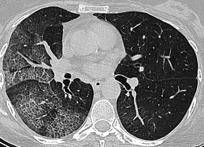
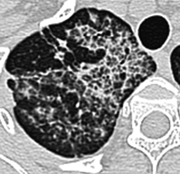
Chronic eosinophilic pneumonia is also characterized by consolidation (Figures 35, 112-113) with subpleural and often apical distribution. The changes may persist for a long period, but generally respond rapidly to cortisone treatment. Approximately half of these patients also have asthma.

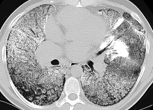
Acute eosinophilic pneumonia is a rare acute febrile illness that may lead to respiratory failure. In radiological imaging terms it is described as edema-like. Cortisone treatment is very effective.

Hypereosinophilic syndrome is a rare multiorgan disease. Eosinophilic infiltrates can be found in the heart, lungs and other internal organs. The HRCT pattern varies from nodules to consolidation.

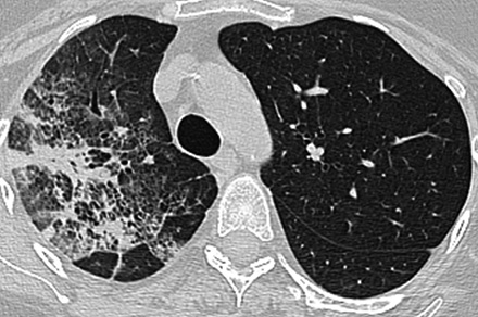
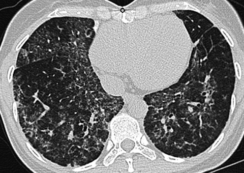
Churg-Strauss syndrome (Figure 114) is a small vessel vasculitis occurring in patients with asthma. The patients have eosinophilia, and lung changes are seen in approximately half of cases. The HRCT pattern varies and includes patchy ground-glass, crazy paving, elements of consolidation and sometimes nodules. The picture is very unspecific, and consequently diagnosis is based on a typical triad of small vessel vasculitis, asthma and eosinophilia.